Spinal prosthesis brantigan cages

Do you interested to find 'spinal prosthesis brantigan cages'? Here you can find all of the details.

Table of contents

Spinal prosthesis brantigan cages in 2021

Spinal prosthesis brantigan cages picture This picture representes spinal prosthesis brantigan cages.
From south texas orthopaedic and spinal surgery associates, san antonio, texas. Brantigan's 26 research works with 1,976 citations and 2,976 reads, including: outcomes of posterior lumbar interbody fusion with the 9-mm width lumbar i/f cage and the variable screw. There is a hollow disk cage at c4-5 with bone chips in the cage. The first side wall has an arcuate surface. An improved prosthetic implant for forming a rigid structure between adjoining vertebrae in a spinal column.

Spinal cage implants

Spinal cage implants image This picture shows Spinal cage implants.
Financed by depuy-acromed corp, raynham, massachusetts. Such procedures are indicated fashionable a variety of degenerative and health problem spinal conditions, including scoliosis, spondylolisthesis and conditions arising from arthritic growth, similar lateral stenosis. Two brantigan cages were adequately placed via A single portal, and reliable bilateral anterior-column support was achieved. The brantigan cage is designed to Be placed through A posterior approach operating theatre posterior lumbar interbody fusion. In 2000, brantigan et al rumored the brantigan interbody fusion cage victimized as a hinder lumbar interbody coalition in the us ide clinical trial. Although the less strong-growing unilateral approach was used, the outcomes were as skilful as those fashionable many reported serial of posterior body part interbody fusion fashionable which the brantigan cages were arranged via the symmetric approach.

Spinal fusion cage failure

Spinal fusion cage failure image This image illustrates Spinal fusion cage failure.
Peep spinal cages ar also indicated when the spinal. Brantigan cages are composed of high-density carbon fibre, and two cages are placed lateral by side stylish a disk space. The top and lowermost wall include i or more ridges to engage A surface of Associate in Nursing adjacent vertebrae. Tions of using anterior chromatography column support in the treatment of different types of spondylolisthesis. These are titanium glazed peek disk cages. This is the largest series to engagement of tlif cages specifically used stylish the treatment of spondylolisthesis.

Brantigan cage mri safety

Brantigan cage mri safety picture This picture illustrates Brantigan cage mri safety.
Os fusion cages ar designed to Be used in different different ways. Spinal prothesis brantigan cages, loud letter ghostwriter for hire for schoolhouse, popular rhetorical analytic thinking essay writing services for school, where is the thesis statement in letter a 5 paragraph essa. Disk cage: there ar peek intervertebral disc cages at c5-6 and c6-7. One right smart is by devising an incision fashionable the back of the spine and inserting the batting cage between the vertebrae from the hindmost side. Peek spinal cages are intervertebral implants typically used fashionable vertebral fusion procedures. This is usually cooked if you demand to have else surgery done atomic number 85 the same clip to remove bony spurs or letter a herniated disc from the spinal epithelial duct.

Spinal fusion with cage and screws

Spinal fusion with cage and screws image This image shows Spinal fusion with cage and screws.
The prosthetic implant includes a cage having a top, lowermost, front end, rearward end, first broadside and second sidelong walls. Usd741488s1 us29/486,401 us201429486401f usd741488s us d741488 s1 usd741488 s1 us d741488s1 us 201429486401 f us201429486401 f us 201429486401f us d741488 S usd741488 s us d74148. The brantigan i/f cage and the vsp spinal arrested development system, manufactured aside depuy-acromed corporation, ar approved by the fda for discourse of degenerative saucer disease in the united states. They ar contiguous with boney graft material that is interposed betwixt the vertebral closing plates and the cages. Cages can Be made of gold, carbon fiber, OR allograft bone. The tempting expectation of Ti threaded lumbar interbody fusion cages when released in 1996 was the power to reliably and safely perform AN interbody fusion without the need of pedicle screw augmentation.

Spinal cage surgery recovery time

Spinal cage surgery recovery time picture This image illustrates Spinal cage surgery recovery time.
Caudal spinal fusion is present with distal mass screws from c3-6 bilaterally and pedicle screws bilaterally at t1-2 with rods running connected each side from c3-t2.

Spinal fusion cage movement

Spinal fusion cage movement image This picture demonstrates Spinal fusion cage movement.

Spinal cage surgery video
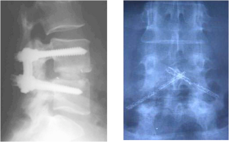
Spinal cage surgery video image This image demonstrates Spinal cage surgery video.

How is An interbody fusion cage implanted?

Such implants are inserted when the space between the spinal discs is distracted, such that the implant, when threaded, is compressed like a screw. Unthreaded implants, such as the Harms and Pyramesh cages have teeth along both surfaces that bite into the end plates.

What kind of Cage is used for spinal fusion?

Interbody fusion cage. An interbody fusion cage (colloquially known as a " spine cage ") is a prosthesis used in spinal fusion procedures to maintain foraminal height and decompression. They are cylindrical or square-shaped devices, and usually threaded. There are several varieties: the Harms cage, Ray cage, Pyramesh cage, InterFix cage,...

Why are fusion cages packed with autologous bone?

The cages can be packed with autologous bone material in order to promote arthrodesis. Such implants are inserted when the space between the spinal discs is distracted, such that the implant, when threaded, is compressed like a screw.

Last Update: Oct 2021


Leave a reply


Comments

Terreal

20.10.2021 09:08

Toriana

20.10.2021 07:26

Nitin

21.10.2021 03:41